
Human anatomy is a complex and fascinating subject. Understanding directional anatomy terms is crucial for high school students studying Anatomy and Physiology or anyone curious about the human body’s structure. For example, these terms are like the compass of anatomy, guiding us through the landscape of the human body. They help us describe the locations of organs, bones, and muscles in a uniform way. In this blog, we’ll explore some of the basic directional terms, such as axis and planes of the body, and concepts like proximal, distal, anterior, and posterior. Grasping these terms is not just about memorizing definitions. It’s important to develop a deeper appreciation for our wonderfully organized and interconnected bodies. So, let’s embark on this journey of anatomical discovery together!
What We Review
The Standard Anatomical Position: The Starting Point of Directional Anatomy Terms
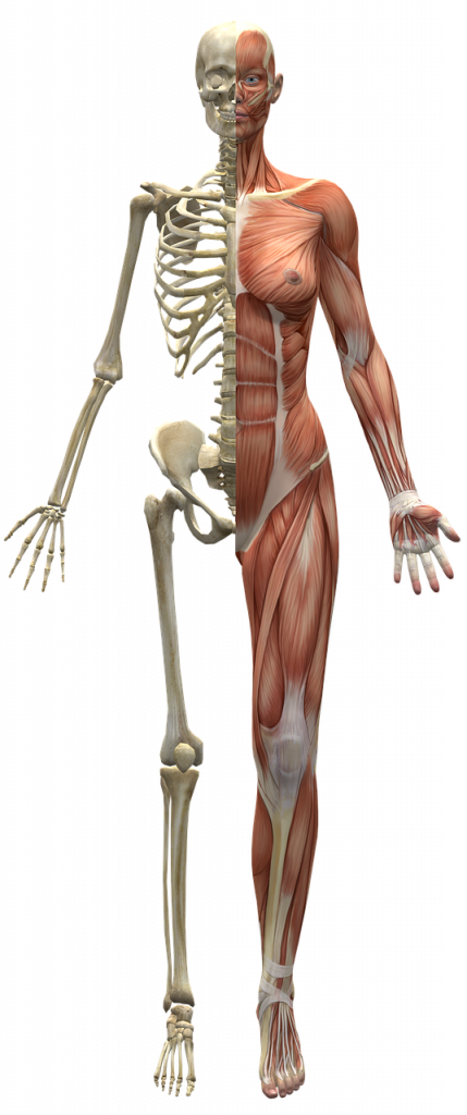
The “standard anatomical position” is the starting point for describing anatomical structures. Picture a person standing upright, facing forward with their feet flat on the ground and close together. Their arms are at their sides. Their palms are facing forward, and their thumbs pointing away from the body. This is “standard anatomical position”. This position is not about how we usually stand or move but serves as a reference for anatomical terminology.
Why is this position so important? It provides a consistent framework for the body. In anatomy, clear communication is crucial. When a doctor talks about pain in the knee, it is important for everyone to understand the exact location being referred to. To have clear communication, medical professionals use standard anatomical terms like ‘anterior’ and ‘posterior’, or ‘proximal’ and ‘distal’. These allow for consistent usage of medical terms, helping patients understand their bodies. This consistency is key to accurate descriptions in healthcare and anatomical research.
In the following sections, we’ll look into more specific terms but always remember: they all relate back to this foundational pose.
Anatomical Planes: Understanding the Body’s Divisions
Anatomical planes are imaginary lines that slice through the body. Planes provide a way to divide and describe the body’s various parts. These divisions are important for medical imaging, surgery, and understanding the human body’s structure. Let’s further examine the three main planes: coronal, sagittal, and transverse.
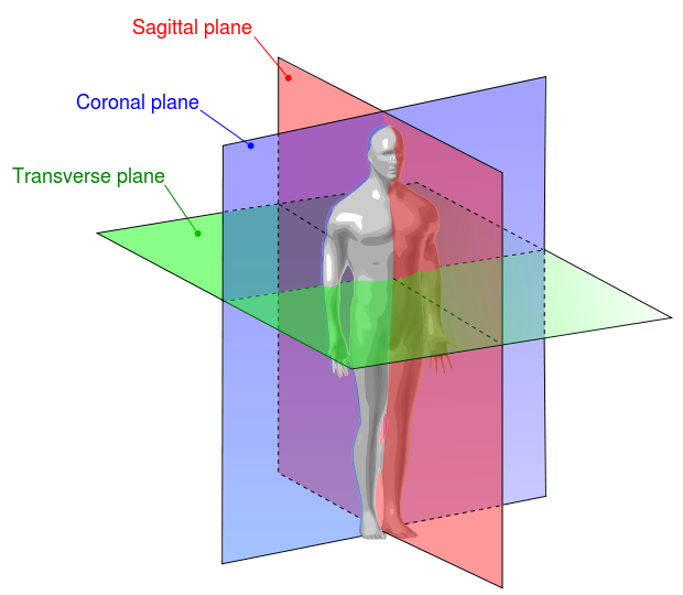
Image source: Wikimedia Commons
Coronal (Frontal) Plane
This plane runs vertically, dividing the body into front and back sections. So, imagine a line passing through the body from one ear to the other. Therefore this line separates the face and chest from the back of the head and back. It’s often referred to in discussions about structures that are towards the front or back of the body.
Sagittal (Longitudinal) Plane
This is another vertical plane that slices the body into left and right parts. Think of a line running down the body’s center, from the top of the head through the navel to the floor. This plane is important when talking about unevenness or movements that occur on either side of the body.
Transverse (Axial) Plane
This plane cuts horizontally, dividing the body into upper and lower halves. Imagine a horizontal line running around the waist, separating the body into top and bottom sections. It’s particularly useful when describing cross-sections or movements that involve the upper and lower parts of the body.
Understanding these planes allows for a more organized approach to studying the body’s anatomy. They provide clear and consistent references for describing the locations and movements of different body parts.
Axis and planes of the body are usually lumped together but are fundamentally different. Continue reading to learn more about the axes.
Axes of the Body: Understanding Movement and Rotation
In anatomy, distinguishing between planes and axes is crucial for understanding how the body moves and rotates. However, the axes and planes of the body can be hard to separate. While a plane is an imaginary flat surface slicing through the body, an axis is an imaginary line around which the body or its parts can spin perpendicular to the plane. So let’s examine the main axes of movement.
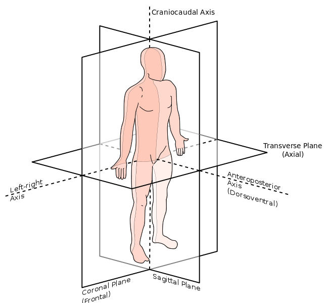
Image source: Wikimedia Commons
Frontal (Left-right) Axis
This axis runs horizontally from left to right through the body’s center. Movements around this axis are seen when the body rotates sideways. For instance, imagine a gymnast performing a somersault. Their body rotates around this left-to-right line.
Anteroposterior (Sagittal) Axis
Positioned from front to back, this axis runs through the body’s center. Movements around the sagittal axis are found in activities like a cartwheel, for example. As a gymnast extends their arms and legs and rotates side-to-side, their body is spinning around this front-to-back line.
Craniocaudal (Vertical) Axis:
This axis goes from the top of the head down through the feet. It’s central to rotational movements that occur in a standing position. For instance, picture a figure skater completing a spin. They rotate around this top-to-bottom line, showcasing movement along the vertical axis.
Generally, understanding these axes helps in analyzing and describing the ways in which different parts of the body move. This provides a more complete understanding of human movement.
Comparative Anatomical Terms: Understanding Body Part Relationships
In anatomy, comparative terms are important for describing the locations of body parts in relation to one another. Let’s explore four sets of these terms to better understand the body’s layout.
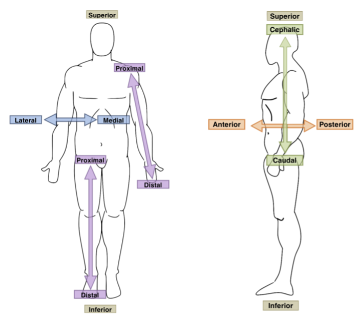
Images sourced from Wikimedia Commons (Image 1) (Image 2)
Inferior vs. Superior
These terms are used to describe higher and lower positions on the body. ‘Superior’ means above or towards the head, while ‘inferior’ indicates below or towards the feet. For example, the nose is superior to the mouth, and the abdomen is inferior to the chest.
Anterior vs. Posterior
These terms describe the front and the back of the body. For instance, the chest is on the anterior side of the body. On the other hand, your shoulder blades are located on the posterior side.
Medial vs. Lateral
These terms relate to the vertical middle of the body. ‘Medial’ means closer to the middle. ‘Lateral’ is used for parts further from the middle. The thumb, for example, is lateral to the little finger, and the nose is medial to the ears.
Proximal vs. Distal
Proximal and distal describe positions along the arms and legs (limbs) compared to where they attach. ‘Proximal’ means closer to where a limb attaches to the body, like the shoulder for the arm or the hip for the leg. Oppositely, ‘distal’ means a point farther from the attachment, like the fingers, compared to the elbow. For instance, the knee is proximal to the ankle but distal to the hip. The terms proximal and distal are used frequently in medicine.
Understanding these terms is not just for class; they’re important in medical settings for clear communication about body part locations and positions. They enable simple, short, and accurate descriptions, which are crucial in medicine.
Applying Directional Terms: Real-World Examples

Now that you’ve learned about different directional anatomy terms let’s see how they are applied in real-world scenarios.
Clinical Descriptions
In a clinical setting, these terms clarify diagnoses and treatment. For instance, a patient might have pain in the distal part of the forearm, indicating the area closer to the wrist. Because of this precise language, healthcare providers can quickly understand and locate the issue.
Physical Therapy and Sports
Understanding these terms is crucial in physical therapy and sports medicine. For example, a therapist might instruct an athlete to strengthen the muscles on the posterior side of the thigh (the hamstrings) to improve performance or recover from an injury. They might also use their knowledge of axis movement and planes in the body to describe joint movement.
Biological Research
In research, describing experiments and anatomy accurately is important. For instance, when studying the effects of a drug on heart health, researchers might examine the anterior part of the heart to understand its impact on a specific heart chamber.
These examples show the practical importance of understanding directional anatomy terms accurately, whether in a healthcare setting, on the sports field, or in a research lab.
Conclusion: Embracing the Language of Anatomy
In conclusion, as we end our journey through the world of directional anatomy terms, it’s important to recognize the impact these terms have on our understanding of the human body. Certainly, these are not just vocabulary words to be memorized. They represent a basic aspect of anatomy and physiology, crucial for students, healthcare professionals, and anyone interested in the workings of the human body.
Because these terms provide a common language, clear communication is possible in many medical situations, from the emergency room to the research laboratory. Furthermore, understanding terms like anterior and posterior, proximal and distal, or axis and planes of the body can help you see the complexity of the human body.
Overall we hope this overview has sparked your curiosity about the design of our bodies. Remember, each term you master is a building block in your foundation of anatomical knowledge, paving the way for a deeper understanding of health, disease, and medicine.


